Why This Matters Now
Medical imaging has long served as a cornerstone of diagnostic medicine, but artificial intelligence is fundamentally transforming its role from passive visualization to active clinical intelligence. Recent FDA clearances and research breakthroughs signal a pivotal shift: AI-powered imaging tools are no longer confined to identifying abnormalities. They now integrate genetic data, predict treatment responses, and guide personalized therapeutic decisions across specialties from oncology to cardiology.
This evolution arrives at a critical juncture for healthcare systems grappling with increasing patient complexity, workforce shortages, and the imperative to deliver precision medicine at scale. As these technologies mature from experimental prototypes to clinically validated tools, they promise to augment—and in some cases fundamentally reshape—how physicians diagnose disease, monitor treatment, and make strategic decisions about patient care. For healthcare organizations and recruiting platforms like PhysEmp, understanding these technological shifts is essential to anticipating workforce needs and supporting the integration of AI-enhanced clinical workflows.
From Detection to Quantification: AI in Brain Metastasis Assessment
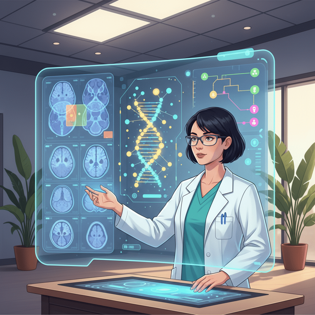
The FDA’s recent clearance of MRI-based AI software for brain metastasis assessment exemplifies how artificial intelligence is moving beyond binary detection tasks toward comprehensive quantitative analysis. Brain metastases represent one of oncology’s most challenging scenarios—these lesions are common in lung cancer, breast cancer, and melanoma patients, yet their accurate measurement and tracking over time demands significant radiologist time and introduces variability in manual assessments.
The newly cleared software employs advanced algorithms to automatically detect, segment, and measure brain metastases across MRI scans. More significantly, it performs longitudinal comparisons, tracking how lesions respond to interventions over time. This temporal analysis capability transforms imaging from a snapshot assessment into a dynamic monitoring system that generates quantitative data to inform treatment decisions about surgery, radiation therapy, or systemic approaches.
The clinical validation studies supporting FDA clearance demonstrated accuracy comparable to or exceeding manual radiologist assessment—a threshold that represents not just technological achievement but practical clinical utility. For oncologists managing patients with metastatic disease, access to consistent, reproducible measurements reduces uncertainty in treatment response evaluation and enables more confident adjustments to therapeutic strategies.
AI imaging tools that provide longitudinal quantification represent a fundamental shift from diagnostic support to treatment monitoring infrastructure. By generating reproducible metrics across time, these systems create data foundations for evidence-based adjustments to cancer therapy that were previously difficult to standardize.
Multimodal Integration: Linking Images, Genes, and Therapeutics
While the brain metastasis tool demonstrates AI’s capacity for enhanced detection and measurement, recent research in cardiovascular imaging reveals an even more ambitious trajectory: the integration of imaging phenotypes with genetic data and drug predictions through AI-powered knowledge graphs. This approach represents a conceptual leap from analyzing single data streams to synthesizing multiple information types into unified clinical intelligence.
Researchers have developed systems that analyze echocardiogram images using machine learning, then connect observed cardiac abnormalities to underlying genetic variants and potential therapeutic interventions. This multimodal integration exemplifies precision medicine’s core promise—tailoring treatment decisions to individual patients based on their unique genetic and clinical profiles rather than population averages.
The knowledge graph architecture enables the AI system to traverse connections between imaging findings, genomic databases, and pharmacological knowledge. When the system identifies a cardiac structural abnormality on imaging, it can suggest associated genetic variants that might explain the phenotype and propose drug candidates with mechanisms relevant to the underlying pathophysiology. In validation studies, these predictions demonstrated accuracy in linking imaging findings to genetic heart disease variants, potentially accelerating diagnosis and enabling more targeted treatment selection.
This capability transcends traditional imaging interpretation. Rather than simply describing what is visible, the AI system generates hypotheses about why abnormalities exist and what interventions might address root causes. For clinicians, this transforms imaging studies from diagnostic endpoints into starting points for genetic investigation and personalized therapeutic planning.
Precision Medicine Infrastructure and Clinical Workflow Integration
The convergence of these technologies—quantitative longitudinal tracking in oncology and multimodal data integration in cardiology—reveals a broader pattern in healthcare AI development. The most impactful applications are those that embed themselves seamlessly into clinical workflows while expanding the analytical scope of existing diagnostic modalities.
For radiologists and cardiologists, these tools don’t replace clinical judgment but rather augment it with capabilities that would be impossible through manual analysis alone. A radiologist cannot realistically measure dozens of brain metastases across multiple time points with perfect consistency, nor can a cardiologist instantaneously connect an echocardiographic finding to relevant genetic literature and drug databases. AI systems excel precisely at these tasks that require processing large data volumes, maintaining consistency, and synthesizing information across disparate knowledge domains.
The clinical value of AI imaging tools lies not in replacing physician expertise but in expanding the analytical bandwidth available during decision-making. By handling quantitative measurement and multimodal data synthesis, these systems allow clinicians to focus cognitive resources on interpretation, patient communication, and strategic treatment planning.
Yet integration challenges remain substantial. Healthcare organizations must address questions of validation, liability, workflow disruption, and clinician training. Physicians need to understand not just how to use these tools but how to interpret their outputs critically, recognizing both capabilities and limitations. The FDA clearance process provides regulatory validation, but clinical adoption requires demonstrating value in real-world settings with diverse patient populations and varying institutional resources.
Implications for Healthcare Workforce and Organizational Strategy
These advances in AI-powered imaging carry significant implications for healthcare workforce development and organizational strategy. As imaging tools evolve from diagnostic aids to precision medicine platforms, the skill sets required of radiologists, oncologists, and cardiologists are shifting. Clinicians increasingly need literacy in AI system capabilities, limitations, and appropriate use cases. They must understand how to integrate AI-generated insights with clinical context, patient preferences, and other data sources.
For healthcare organizations, strategic questions emerge about technology adoption timing, vendor selection, and workflow redesign. Early adoption of validated AI tools may provide competitive advantages in care quality and efficiency, but requires investment in infrastructure, training, and change management. Organizations must also consider how these technologies affect staffing models—if AI systems handle routine quantification and preliminary analysis, how does that reshape radiologist workload and subspecialty demand?
Recruiting platforms focused on healthcare, such as PhysEmp, must anticipate these workforce evolution patterns. Job descriptions increasingly specify familiarity with AI-assisted diagnostic tools. Institutions seek candidates who combine clinical expertise with technological adaptability. Understanding these trends enables more effective matching between healthcare organizations navigating AI integration and professionals equipped to thrive in technology-augmented environments.
Longer-term, the proliferation of AI imaging tools that generate structured, quantitative data creates opportunities for learning health systems where clinical outcomes continuously inform algorithmic refinement. As these systems accumulate data across diverse patient populations, their predictive accuracy for treatment response and genetic associations should improve, creating virtuous cycles of enhanced precision medicine delivery.
The trajectory is clear: medical imaging is transforming from a primarily visual diagnostic modality into a multimodal analytical platform that integrates genetics, pharmacology, and longitudinal outcome tracking. Organizations and professionals who recognize and adapt to this shift will be best positioned to deliver the next generation of precision medicine.
Sources
FDA Clears MRI-Based AI Software for Assessment of Brain Metastases – Diagnostic Imaging
AI-powered knowledge graph links heart images to genes and drug predictions – Medical Xpress
")

")


